Copyright – disclaimer – foto bronnen
AbortusInformatie.nl besteedt de grootst mogelijke aandacht voor en zorg aan de gegevens en informatie die op deze site te vinden is. Desondanks is het mogelijk dat er in de website onjuistheden en/of onvolkomenheden voorkomen. Hoewel de grootste zorg is besteed, kan de organisatie niet aansprakelijk worden gesteld voor de juistheid van alle informatie. Alleen ter informatie en voor onderwijs doeleinden.
Wij aanvaarden geen aansprakelijkheid voor schade als gevolg van deze onjuistheden en/of onvolkomenheden, noch voor problemen die worden veroorzaakt door het gebruiken of verspreiden van deze informatie.
AbortusInformatie.nl aanvaardt evenmin aansprakelijkheid voor geleden verlies of gederfde inkomsten welke voortkomt uit het gebruik of verspreiden van de informatie, dan wel voortkomt uit technische gebreken. Bezoek aan onze websites, alsmede gebruik en downloaden van gegevens en informatie geschiedt geheel op risico van de gebruiker.
Informatie op websites waar naar wordt verwezen of gelinkt, wordt slechts incidenteel door ons geanalyseerd. Wij geven geen garanties met betrekking tot de inhoud en betrouwbaarheid van deze sites, noch wordt aansprakelijkheid aanvaard voor direct of indirect geleden schade welke hieruit voortkomt. Door te linken wil niet zeggen dat wij het eens zijn met die bron.
Beelden gebruiken van AbortusInformatie.nl en Center for Bio-Ethical Reform CBR is alleen toegestaan na overleg, en met correcte bronvermelding, inclusief:
CBR in NL: AbortusInformatie.nl
Deze website is niet bedoeld om medisch advies, diagnose of behandeling te verstrekken.
Hoewel de grootste zorg is besteed aan het samenstellen van deze website, kan de organisatie niet aansprakelijk worden gesteld voor de juistheid van alle informatie.
Wij zijn niet aansprakelijk voor eventuele schade door eventuele onvolledigheden, onjuistheden of vermeende adviezen op deze site. Alleen ter informatie en voor onderwijs doeleinden.
De inhoud van deze website is niet bedoeld als vervanging van de geïndividualiseerde raad van een arts.
This website is not intended to constitute medical advice or replace the individualized counsel of a doctor.
Live image source
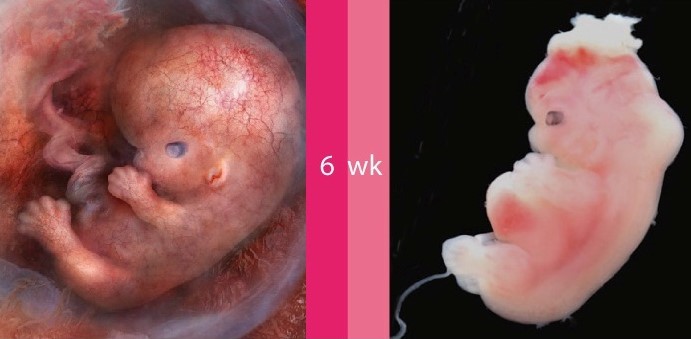
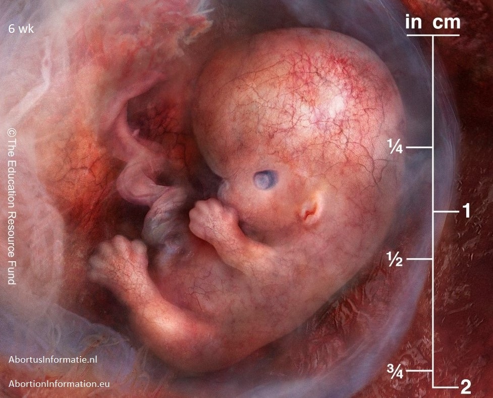
CBR’s embryo and fetus pictures are derived from many smaller images “stitched” together in much the same manner NASA uses to combine satellite photo “tiles” to form a large “mosaic.”
Endoscopes
The Center for Bio-Ethical Reform’s (CBR) human embryo and fetus imagery was initially derived by teams of physician researchers and clinicians employing endoscopy (and its subsets, embryoscopy and fetoscopy) to diagnose and treat prenatal disorders in utero. Endoscopes are medical imaging devices which permit the minimally invasive, high resolution observation of tissues inside the human body. At the distal end of these instruments is an objective lens designed for imaging. At the proximal end is an eyepiece, or sensor, which enables viewing.
How Endoscopes Work
These scopes generally consist of a tube which encloses a relay lens system (in rigid endoscopes) or a fiber bundle (for fiber-optic, or flexible, endoscopes) for illumination and to transmit an image from the objective lens inside the body to the proximal end outside.
Said differently, endoscopes use optical elements to direct light to the area sought to be illuminated and transmit the resulting image to the eye or detector. Rigid endoscopes generally offer superior resolution or magnification. But an endoscope’s objective lens is only approximately 1/5 of an inch in diameter, and this relatively small size substantially narrows the observer’s field of view (even with the addition of supplemental lenses such as “negative” or “prism” optics, etc.)
Constraints of Endoscopes
This limitation is further compounded by the need to use the scope in very confined spaces, with only short distances separating the objective lens from the anatomical structures being imaged. As a consequence, only a small segment of the embryo or fetus is observable at any point along the timeline of the scan. An endoscope’s construction must also accommodate frequently conflicting design considerations. The resulting compromises can involve not only fields of view, but depths of field (meaning thickness of the plane of focus) and image illumination and magnification, as well as distortion issues (i.e., stretched or compressed perspective), etc.
Work-Arounds to the Constraints of Endoscopes
So to depict a high quality, single image of the entire embryo or fetus, large numbers of smaller, more detailed pictures must be joined together in a manner suggestive of the process by which puzzle pieces are assembled to form a completed picture.
This technique employs a complex proprietary process which combines segmental scans to create a final composite image. The resulting picture is digitally adjusted to preserve each segment’s original colour, resolution, contrast, illumination, etc. Technicians also correct for vignetting (image degradation or loss at the periphery of the frame).
Magnetic Resonance Imaging and Ultrasound
Our process resembles the image enhancement process described in the British medical journal Lancet which recently published a prenatal magnetic resonance imaging (MRI) study involving the creation of 3D pictures to diagnose and treat congenital heart problems afflicting fetuses still in the uterus. The BBC reports that “A series of 2D pictures of the heart are taken from different angles using an MRI machine” to image the fetus.
The story explains that “Sophisticated computer software pieces the images together, adjusts for the beating of the heart and builds … [a] 3D image of the heart.” A paediatric cardiologist describes the resulting 3D images as “beautiful.”
This MRI research is part of a fetal diagnostic project which is also exploring scans using “four ultrasound probes at the same time – current scans use one – to get a more detailed picture.” This process produces a more wholistic composite image.
NASA Composite Imagery (“Mosaic”)
CBR’s imaging process is also conceptually similar to the technologies used by the National Atmospheric and Space Administration (NASA) to produce wide-area satellite images of the earth’s surface. Until the launch of the Deep Space Climate Observatory Satellite (DSCOVR), which now orbits one million miles from the surface, NASA had no camera positioned sufficiently far from earth to capture the entire globe in a single photograph. As previously noted, an endoscope’s objective lens must also operate too near to an embryo or fetus to permit its whole anatomy to be imaged in a single frame. This is the same constraint which complicates the capture of satellite imagery. Previous pictures of the earth could, therefore, only be created using digital stitching technology to make one large composite image from smaller segments. Scientists sometimes describe this final image (or “data set”) as a “mosaic,” comprised of many individual “tiles.”
Hybrid Imagery (Multi-Media)
A satellite picture can also be augmented by aerial photography (cameras on aircraft platforms) to improve image resolution. Hybrid images of this sort can be created by superimposing black-and-white imagery (for still higher resolution) over colour pictures of the same area, the latter to optimise chromic (colour) fidelity.
The scientific press, for instance, reports that the Landsat Image Mosaic of Antarctica (LIMA) “combined over one thousand precise, calibrated satellite images with other data from the continent’s surface to create a single picture of the entire continent.” The high magnification factor (think telephoto lenses which enlarge image features) of each of these puzzle pieces yielded a composite picture depicting more detail than would have been visible in a single photo shot with a wide-angle lens.
Applications for “Mosaicking”
NASA uses this mosaicking process to image celestial bodies of nearly every description. The Juno spacecraft made composite images of Jupiter; InSight of Mars; Cassini of Saturn; and Hubble of the Sombrero Galaxy.
So an exquisite depiction of a challenging subject, whether prohibitively small or large, near or far, may involve vastly more complexity than meets the eye of the casual observer.
Photo authenticity
Center for Bio-Ethical Reform CBR is waar wij mee gelieerd zijn, maar geheel zelfstandig opereren. Cbrinfo.org
The history of social reform teaches us the power of images: in raising awareness of an issue, changing public opinion, and bringing about social reform.
https://www.abortionno.org/international-resources/
https://www.abortionno.org/prenatal/
https://www.abortionno.org/abortion-photos/
https://www.abortionno.org/lawsuits/verifying-photograph-authenticity/
Images by Center for Bio-Ethical Reform of aborted babies with recognition / authentication by an abortionist, pathologist, certified by notary with stamps – witnessed.
CBR is preparing to take legal action against pro-abortion defendants who falsely accuse CBR of fraudulently altering pictures of aborted embryos and fetuses. We will present affidavits from our photographers and certifications of authenticity from technical experts who have examined our original negatives, transparencies and videotape. We will also rely on the expert testimony of physicians who have formerly practiced abortion medicine.
One example of this type of authentication is contained in a letter we recently received from Anthony P. Levatino, M.D., J.D. Dr. Levatino is both a physician and attorney and he says the following:
I, the undersigned, having performed induced abortions earlier in my career, have examined the photos depicting the aborted human embryos and fetuses used by The Center For Bio-Ethical Reform in their public education projects (http://www.abortionNO.org). It is my professional opinion that the photos depict aborted human embryos and fetuses and that the depicted aborted human embryos and fetuses are accurately captioned as to age, in weeks since fertilization.
Anthony P. Levatino, M.D., J.D.
Compare our size references to the textbooks’ prenatal development scales. We encourage anyone who wishes to verify the truth to examine the embryology photos in the following academic texts, used in medical schools the world over:
The first source is the authoritative, William’s Obstetrics, 20th Edition, Gary Cunningham, MD, Paul C. MacDonald, MD, Norman F. Gant, MD, Kenneth J. Leveno, MD, Larry C. Gilstrap III, MD, Gary D. V. Hankins, MD & Steven L. Clark, MD (Copyright 1997 by Appleton and Lange, A Simon & Schuster Company) beginning at page1026, Table 44-4, “Predicted Menstrual Age (MA) in Weeks From Crown-Rump Length (CRL) Measurements (in Centimeters)”
CBR’s physicians also rely on a highly regarded book called The Color Atlas of Clinical Embryology, 2nd Edition, Keith L. Moore, T.V.N. Persaud & Kohei Shiota (Copyright 2000 by W. B. Saunders Company) at page 49, Table 2–1, “Criteria for Estimating Developmental Stages in Human Embryos.” This reference contains age to crown/rump length relationships, etc. from 19-21 days post-conception through 56 days (the end of the embryonic period). At page 52, Table 3-1, you will find criteria for establishing age through the fetal period, including age-crown/rump length relationships from 9 weeks through 38.
Dr. Moore has also published a popular text entitled The Developing Human, Clinically Oriented Embryology, Keith L. Moore & T.V.N. Perasud, 6th Edition, (Copyright 1998 by W.B. Saunders) which can be used for aging unborn babies by turning to page 91 (see also, tables on pages 4 through 7).
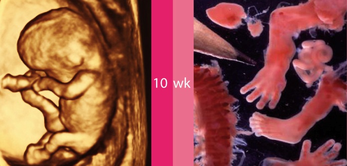
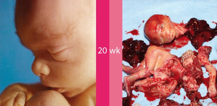
We don’t ask anyone to take our word for anything. Doubters can go to our Website and hold these textbook illustrations next to the corresponding photos on our signs. When measuring our images, bear in mind that many of the first trimester aborted babies we show have been torn apart by the termination procedure. The shredded pieces of their bodies have only been loosely reassembled for photographic purposes. Abortion stretches, tears and otherwise distorts the soft tissue of these very small unborn babies.
Of course, no amount of evidence will satisfy dogmatic pro-aborts. They wouldn’t acknowledge the legitimacy of any abortion photos, even if they took the pictures themselves — of abortions performed in their very presence. But for sincere questioners, those who inquire in good faith, the foregoing sources will quiet every reasonable concern.
We are sometimes asked how and from what sources we have compiled our huge library (perhaps the largest in the world) of aborted baby photos and video. Understandably, the terms of our acquisition agreements prohibit the disclosure of that information. If we must divulge this information pursuant to the lawsuits we are preparing, we will do so “in camera,” which means in a judge’s chambers, off the record, so the information will be sealed and never made public. It doesn’t take much analytical ability to guess why. We are prepared, however, to say that we reject civil disobedience on tactical grounds (and violence on moral grounds) so we use only lawful means to acquire imagery.
Copyright anderen
Besides being the Dutch affiliate of CBR Center for Bio-Ethical Reform (cbrinfo.org), which includes being allowed to use their material with our own name on them, we have also been granted permission to use images from other sources.
For example: L.Caustic.
He wrote us in 2019 reaffirming the earlier permission he had given us to be allowed to use his photo’s.
He is the owner of images of miscarried or ectopic pregnancy. These images – although the child has sadly died naturally – give impressions of how babies develop in the womb.
He writes that all of the specimens examined were patients admitted to a USA hospital, and were spontaneous losses (miscarriage) or were ectopic pregnancies. All were examined at the request of the attending physician for Pathological evaluation.
All these images have been posted in an anonymous fashion so that no patient identication is present in the image or as a part of the image name. He may not provide information that breaks HIPAA rules – Health Insurance Portability and Accountability act, USA (i.e. privacy).
Furthermore, many images were made by Maria, who further wishes to remain anonymous, and gave us copyright of these photo’s.
Also, many paintings were made especially for us by Irene Langeveld, who gave us copyright of these paintings for our use.
Other people have also painted for us, and given us the rights to use their work under our own name.
Some mothers have given us copyright use of their own photo’s of lost children, either through abortion (their child they saw after they used the abortion pill), miscarriage (so we can see what a baby at a certain age looks like), or their ultrasound image, while others gave us permission to use their photo’s of their born children, after changing their mind due to information/interaction with us, and/or the development of the their children in the womb whilst growing.
We are very grateful for all those who have shared and given us permission to share these amazing images with the world, to educate on the beautiful humanity of children growing in the womb.
AbortusInformatie.nl is gelieerd aan CBR Center for Bio-Ethical Reform
Leeftijd meestal na conceptie, niet amenorroe. Als je amenorroe wilt, voeg dan 2 weken toe.
Fotocredit
Center for Bio-Ethical Reform CBR
Ehd.org
Eigen werk AbortusInformatie.nl
https://www.facebook.com/ScienceNaturePage/videos/995057113959880/ Hashem Ghali
en.wikipedia.org/wiki/File: Early_human_embryos.png ekem
Prof. Andrzej Skawina Ph. D., M. D.
Abortion Pill Reversal.com
E. Euthman
Eke
Priests for Life
Mindy Danison Liveaction.org Anabelle
Live action news
Suparna Sinha
L. Caustic